FIGURE 7.
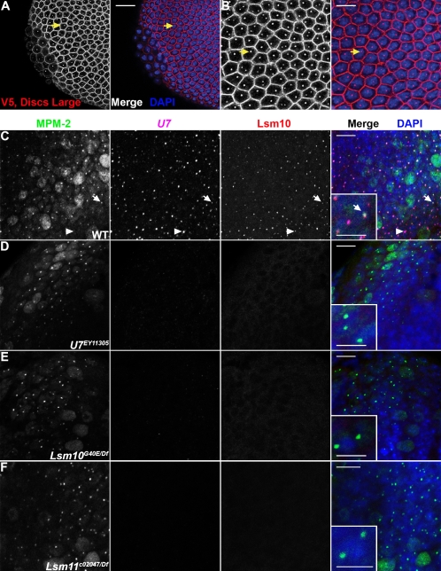
The U7 snRNP formed in an Lsm11 mutant does not localize to the histone locus body. (A) Stage 5 Lsm11c02047, P[V5-Lsm11+] homozygous embryos were stained with anti-Discs Large antibodies, to visualize cell boundaries, and anti-V5 antibodies (left panels, both red in merge). Anti-mouse secondary antibodies were used to simultaneously detect V5-Lsm11 and Discs Large. Embryos were also stained with DAPI (blue in merge). Note that V5-Lsm11 localizes to one or two nuclear foci just like endogenous Lsm11. Arrows indicate the same cell in (A) (20 μm scale bar) and (B) (10 μm scale bar). (C–F) Brains dissected from w1118, U7EY11305, Lsm10G40E/Df, and Lsm11c02047/Df third instar larvae were stained with MPM-2 (first column; green in merge), hybridized with a fluorescent probe recognizing U7 snRNA (second column; magenta in merge), anti-Lsm10 antibodies (third column; red in merge), and DAPI (blue in merge). Arrows indicate a histone locus body that contains MPM-2 antigen(s), U7, and Lsm10. (Insets) A higher magnification view. Arrowheads indicate a histone locus body lacking MPM-2 staining. This nucleus is likely not within S phase, and therefore lacks the Cyclin E/Cdk2 activity necessary to produce the MPM-2 epitope. Note that both U7 and Lsm10 are undetectable in histone locus bodies marked by MPM-2 staining in both Lsm10 (E) and Lsm11 (F) mutant brains. Bar, 10 μm (main panels) and bar, 5 μm (insets).

