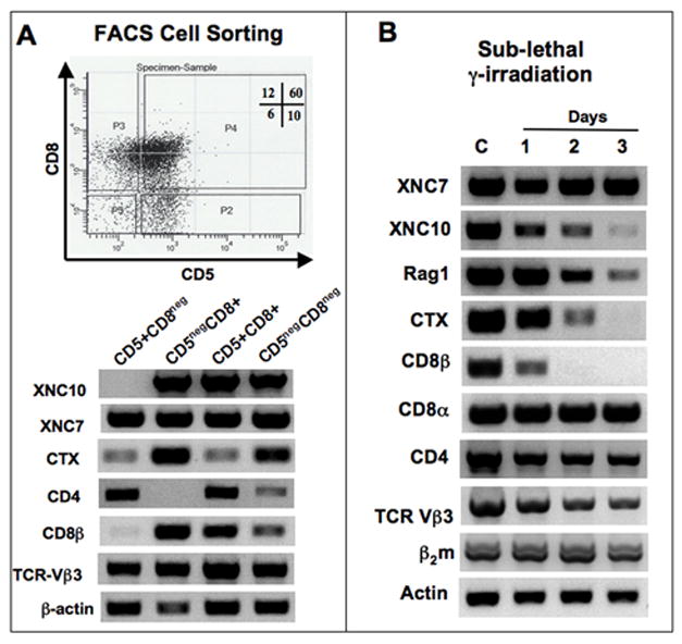
Figure 9. Expression of XNC10 by radiosensitive subsets of larval CD8+ thymocytes, but not by putative mature CD4+ thymocytes.

(A) RT-PCR analysis of thymocytes pooled from a population of 50 pre-metamorphic tadpoles and sorted by FACS according to their expression of CD5 and CD8 surface markers. Upper panel, dot plot of CD5 and CD8 expression with the gate used to sort each population: P2 (CD5+CD8neg), P3 (CD5 negCD8+), P4 (CD5+CD8+) and P5 (CD5negCD8neg). Cells from each population were used for RT-PCR using primers specific for XNC7, XNC10, CD4, CD8β, CTX, TCR-Vβ3, as well as β2m and β-actin as controls. (35 cycles for all genes except, CD8β: 40 cycles, β-actin: 25 cycles). (B) RT-PCR analysis of total thymocytes from pre-metamorphic tadpoles untreated, or 1 to 3 days following sub-lethal γ-irradiation (10 Gray).
