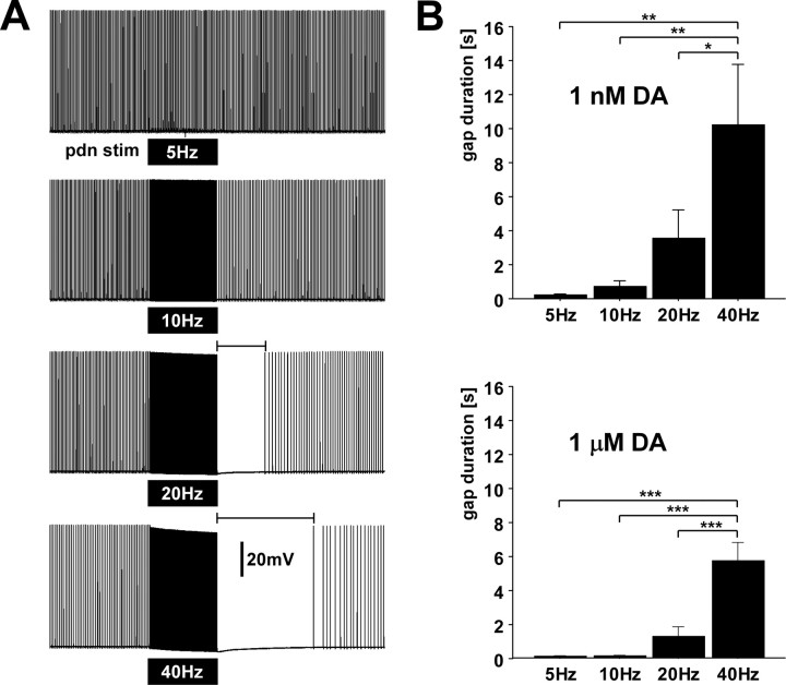
Figure 10.
High frequency spiking suppresses dopamine elicited peripheral spike initiation. A, PD axon recording in the absence of centrally generated activity and the presence of 1 μm dopamine. Black boxes indicate timing of 10 s trains of pdn stimulations at different frequencies. stim, Stimulation. B, Quantification of the interval from the last stimulus to the first spike afterward for different stimulus frequencies in both dopamine concentrations.

