
Figure 11.
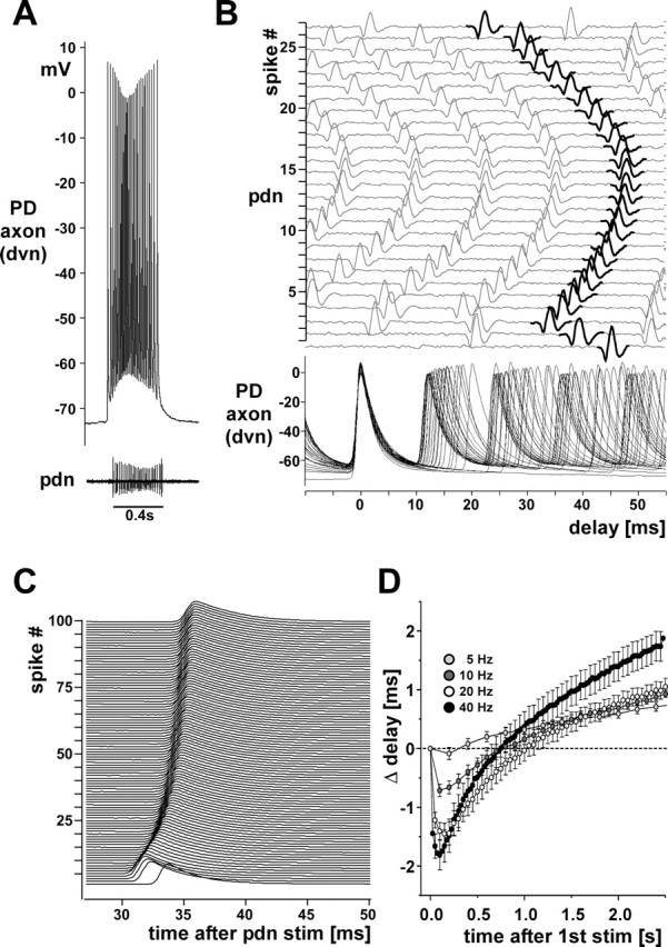
Changes in conduction delay. A, A single burst from a PD axon recording in an experiment in which the other PD neuron signal was missing on one of the pdns. B, Multiple sweeps of the recordings shown in A. Traces are aligned at the peaks of the intracellular spikes. Sweeps of the pdn recording are plotted with an offset and in order, with the extracellular spikes that correspond to the intracellular ones shown in black. Note the change of delay over the course of a single burst. C, Offset multiple sweeps from an intracellular PD axon recording during a 40 Hz extracellular stimulation of the pdn. One hundred stimuli are shown aligned at the pdn stimulus time. Delay initially decreases and then increases. D, Plot of the change in conduction delay over 2.5 s of pdn stimulations with 5, 10, 20, and 40 Hz (n = 28). stim, Stimulation.