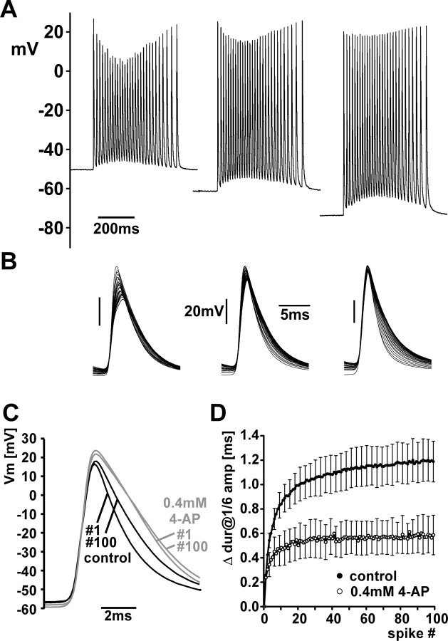
Figure 5.
Changes in spike shape are likely due to channel inactivation. A, Bursts from a PD axon recording at three different interburst membrane potentials set by current injection through a second electrode. The change in spike amplitude is more pronounced at more depolarized potentials. B, Multiple sweeps from the bursts shown in A. The change in spike duration is more pronounced at more hyperpolarized potentials. C, The 1st and 100th spike from a PD axon recording in response to 20 Hz stimulation of the pdn, in control saline and 0.4 mm 4-AP. In 4-AP, duration was increased but did not change as much between 1st and 100th spike. Vm, Membrane potential. D, The increase of spike duration as a function of spike index in control saline (black) and 4-AP (gray). dur, Duration; amp, amplitude.

