
Figure 2.
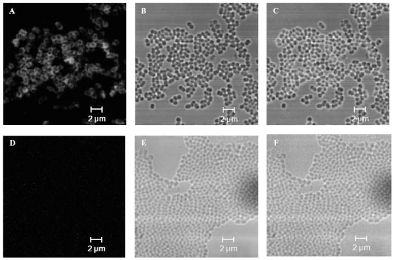
Representative confoal microscopy images showing that V-PA immunogen was loaded on GEM vaccine particle. Staining was done using anti-c-Myc mAb and QD-conjugated antimouse IgG as described in Section 2 and Supporting Information. (A), (B), and (C) are fluorescent, DIC, and merged fluorescent- differential interference contrast (DIC) images, respectively for GEM particle vaccine containing V-PA antigen. (D), (E), and (F) are fluorescent, DIC, and merged fluorescent-DIC images, respectively, for the control sample, GEM particles incubated with Y. pestis V antigen without PA, respectively. No fluorescence was observed for another control, GEM particles incubated with PBS (data not shown).