
Figure 1.
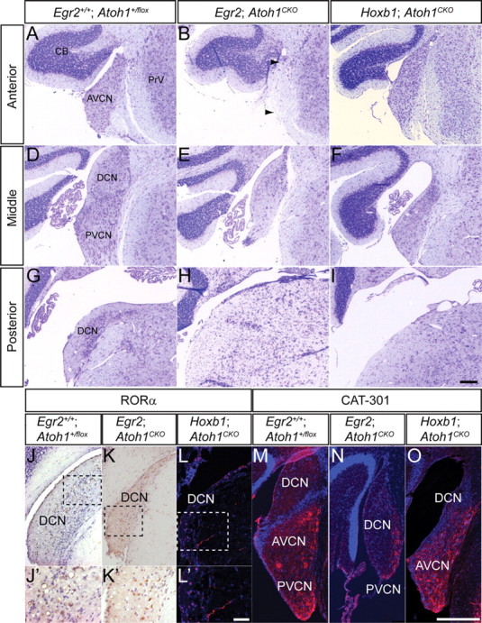
Different subnuclei of the CN are disrupted in adult Egr2; Atoh1CKO and Hoxb1; Atoh1CKO mice. A–I, Cresyl-violet-stained sections of CN from wild-type (A, D, G), Egr2; Atoh1CKO (B, E, H), and Hoxb1; Atoh1CKO (C, F, I) animals at anterior (A–C), middle (D–F), and posterior (G–I) levels. The AVCN is most severely affected in Egr2; Atoh1CKO mice, although the auditory nerve fibers are preserved (B, arrows). The PVCN and DCN are most severely affected in Hoxb1; Atoh1CKO animals. J–L, RORα immunostaining of DCN from wild-type (J, J′), Egr2; Atoh1CKO (K, K′), and Hoxb1; Atoh1CKO (L, L′) animals. RORα-positive cells, which are present only in the DCN, are seen in all three genotypes. J′–L′, Higher-magnification views of boxed areas in J–L, respectively. M–O, Cat-301 immunostaining is present in the VCN of wild-type (M), Egr2; Atoh1CKO (N), and Hoxb1; Atoh1CKO (O) animals. Scale bars: A–I, M–O, 200 μm; J–L, 100 μm; J′–L′, 12.5 μm.