Figure 2.

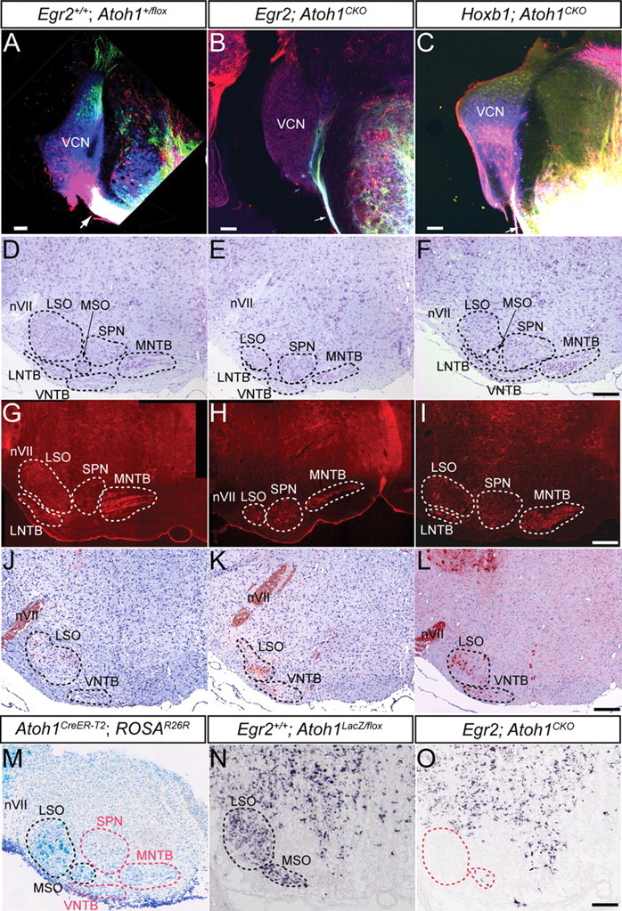
The brainstem AAN and pathways are disrupted in Egr2; Atoh1CKO and Hoxb1; Atoh1CKO mice. A–C, Lipophilic dye injections into the acoustic stria at P18 retrogradely label the CN of wild-type and Hoxb1; Atoh1CKO (A, C) but not Egr2; Atoh1CKO (B) animals. NeuroVue dyes were injected into the ventral acoustic stria near the facial nerve (red) and trigeminal nerve (blue) and into the dorsal and ventral acoustic striae between the other two injections (green). Note the massive filling of the ventral acoustic stria and retrograde filling of DCN (green cells) and VCN (blue and red cells) in wild-type (A) and Hoxb1; Atoh1CKO (C) mice. The caliber of the ventral acoustic stria is much smaller in Egr2; Atoh1CKO animals (compare single arrows in A–C), showing only fibers projecting into the ventral cochlear nucleus that may represent inferior colliculus, superior olive, or trigeminal fibers known to reach the CN in mice. The ventral acoustic stria is also reduced in size in Hoxb1; Atoh1CKO mice (arrow), but retrogradely labeled neurons are still found throughout the CN. For the remaining panels, sections from similar anteroposterior levels of the brainstem are presented for each of the genotypes. D–F, Cresyl violet stain of AAN in 7-week-old wild-type, Egr2; Atoh1CKO, and Hoxb1; Atoh1CKO mice. The nuclear subdivisions are outlined and labeled. The LSO, MNTB, MSO, and VNTB are reduced in size in Egr2; Atoh1CKO animals (E), whereas only the LSO appears to be affected in Hoxb1; Atoh1CKO animals (F). G–I, Cat-301 immunostaining of accessory auditory nuclei shows loss of neurons from the LNTB, LSO, and MNTB of Egr2; Atoh1CKO animals and cell loss from only the LSO of Hoxb1; Atoh1CKO animals. J–L, ChAT immunostaining of brainstems from wild-type, Egr2; Atoh1CKO, and Hoxb1; Atoh1CKO mice confirms that, in all genotypes, neurons in the LSO and VNTB that project in the olivocochlear bundle are present. Note the increased packing density and different positions of these neurons in the LSO of Egr2; Atoh1CKO and Hoxb1; Atoh1CKO compared with wild-type animals. M, X-gal stain of AAN from E18.5 Atoh1CreER-T2 embryo whose pregnant dam was injected with tamoxifen at E10.5. Labeled cells are found in the developing LSO and MSO (black dotted lines). AAN without labeled cells are designated by red dotted lines. N, O, VGLUT2 in situ hybridization of brainstem sections from E18.5 wild-type (N) and Egr2; Atoh1CKO (O) embryos. The wild-type distribution of VGLUT2 mRNA in the AAN is virtually identical to the X-gal expression pattern in M. Only a few VGLUT2-expressing cells are present in the LSO and MSO of the Egr2; Atoh1CKO embryo, suggesting a primary effect of Atoh1 deletion. Red dotted lines in O show the expected position of these cells. nVII, Seventh nerve. Scale bars: A–C, 100 μm; D–O, 200 μm.
