
Figure 5.
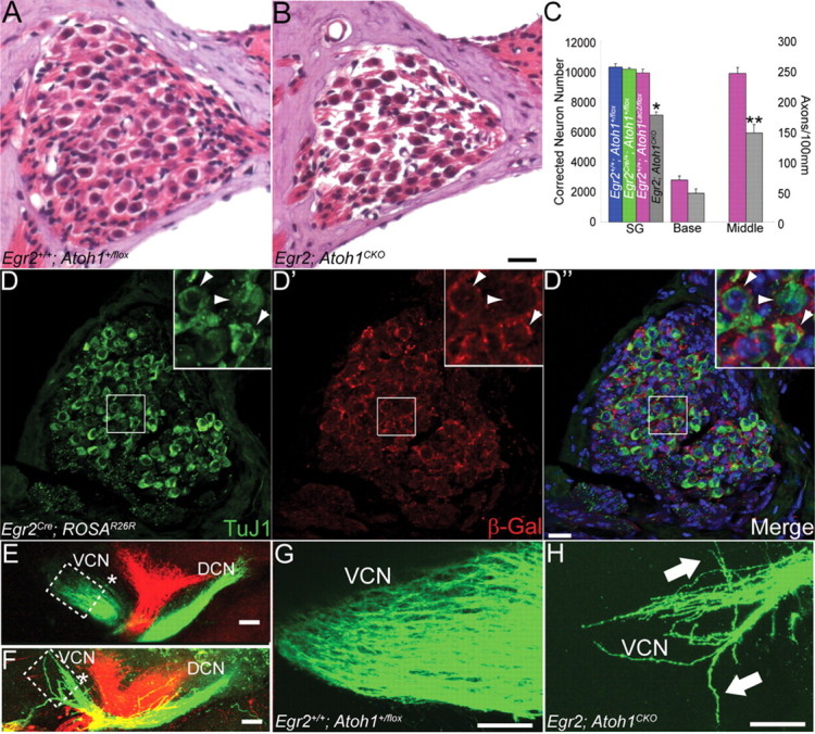
SGN and auditory nerve axon number are decreased in Egr2; Atoh1CKO animals, and auditory nerve axons show pathfinding errors in the CN. A, B, Hematoxylin and eosin-stained spiral ganglion of 6-week-old wild-type (A) and Egr2; Atoh1CKO (B) animals. Note the decreased packing density in the Egr2; Atoh1CKO spiral ganglion. C, Total spiral ganglion neuron number and auditory nerve axon number at the base and middle of the cochlea. Error bars show SEM. All three populations are reduced by ∼30% in Egr2; Atoh1CKO animals relative to wild-type (t test, *p = 0.01, **p = 0.004). D–D″, Spiral ganglion sections from an adult Egr2Cre; ROSAR26R animal immunostained for TuJ1 and β-Gal. Insets show higher-magnification views of the boxed region in each panel. Arrowheads denote TuJ1-positive neurons that are surrounded by Egr2-lineal cells. E–H, Lipophilic dye injections into the apex (green) and base (red) of P0 wild-type (E, G) and Egr2; Atoh1CKO (F, H) cochleae label auditory nerve projections into the VCN and DCN. The VCN projections, particularly those from the apex of the cochlea, are much thinner in Egr2; Atoh1CKO mice, and fibers aberrantly defasciculate and travel in abnormal anterior and posterior directions (H, arrows). Boxes in E and F delineate areas shown in G and H, respectively. * denotes the body of the VCN, which is much smaller in the Egr2; Atoh1CKO CN. Projections to the DCN in Egr2; Atoh1CKO animals are present and appear to be less affected than their VCN counterparts. Scale bars: A, B, D–D″, 20 μm; E–H, 100 μm.