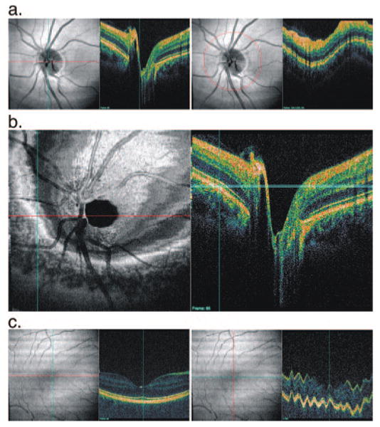
Figure 1.

(a) SD-OCT sample images showing (left) horizontal slice and (right) circular slice. (b) Conventional C-mode view of human retina obtained by prototype SD-OCT unit (501 × 180 × 1024 samplings in a 6 × 6 × 1.4-mm region). C-mode image (left) shows a concentric circular texture and a distorted view of the target layer structure that are caused by slicing multiple different layer structures in the retina with a straight line (light blue band, right). (c) A 3D SD-OCT data set that may appear to have minimal eye motion on viewing of a horizontal section (left) shows significant axial eye motion along a vertical section (right).
