
Figure 3.
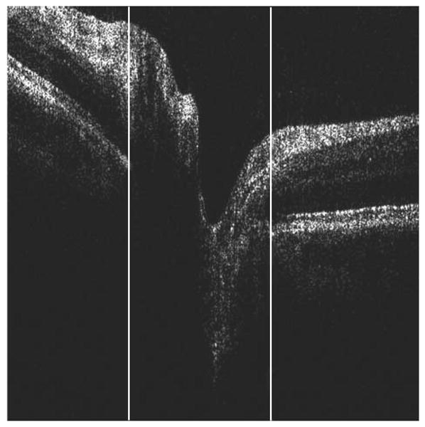
Marking of retinal pigment epithelium (RPE) edges on the cross-sectional optical coherence tomography image. An observer specified the left and right sides of the RPE edges by dragging white lines, as shown. Only the x-coordinate was recorded because the projection of the disc margin location on the en face image was independent of the depth of the RPE termination. In cases in which no RPE edges were observed, the observer flagged the frame as “no edges.”