
Figure 7.
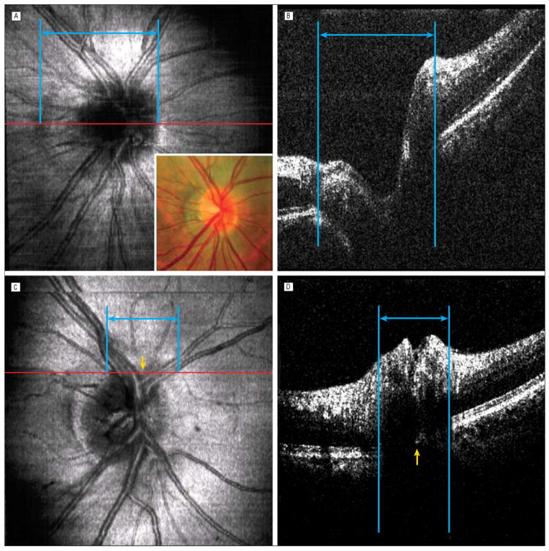
Effects of peripapillary atrophy and vessel shadowing. A and B, Detecting retinal pigment epithelium (RPE) edges on cross-sectional images (B, the frame corresponding to the red line on the optical coherence tomography [OCT] fundus image, A) can be difficult with peripapillary atrophy and major vessel shadowing. B, Because of a weak signal and the sharp kink in the layer representing the RPE-choriocapillaris complex, where the peripapillary atrophy was observed, the left RPE edge was defined erroneously at the margin of the peripapillary atrophy. Because of multiple large vessels at the region (blue lines and arrows), the disc margin identified on the OCT fundus image (C, yellow arrow) was obscured by the shadowing artifact on the cross-sectional image (D, yellow arrow) (the frame corresponding to the red line on the OCT fundus image, C). Corresponding blue lines and arrows indicate where OCT experts defined the RPE edges.