
Figure 5.
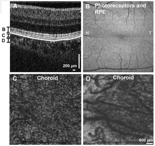
En face visualization of the outer retina and choroid in the macular region by axial integration of 3D OCT data. (A) Cross-sectional image showing the contour of Bruch's membrane (white line), and the axial integration ranges for (B–D). Shown are en face images of (B) the photoreceptors and RPE; (C) the choroid showing predominantly smaller vessels; (D) of the choroid showing predominantly larger vessels. Beam diameter at cornea: 1.4 mm. 512 × 850 axial scans.