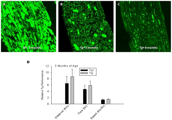
Figure 7. Neural phenotype of VMD2-IL-10 transgenic mice.
A, Tg− sciatic nerve sections are strongly reactive with the SMI-31 antibody, indicating normal neurofilament phosphorylation and phenotype. B, Sciatic nerve sections from 3 month-old Tg+ mice demonstrate decreased reactivity with SMI-31, indicating a decreased loss of neurofilament in the early stages of disease prior to observable levels of cellular infiltration. C, Tg+ sick sciatic nerve sections, however, demonstrate even less reactivity with SMI-31, indicating dephosphorylation of neurofilaments and neural loss correlating with cellular infiltration. D, Voluntary wheel running (VWR) experiments with Tg+ mice (n = 3) before cellular infiltration and disease onset (3-months old) reveals no loss of motor function (distance p = 0.320; time p = 0.341; speed p = 0.576) prior to macrophage infiltration compared to age-matched Tg− littermates (n = 3).

