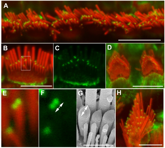
Figure 1. Pan-twinfilin localizes to the tips of shorter stereocilia.
Confocal images showing the distribution of pan-twinfilin in stereocilia bundles. Actin filaments were counterstained with rhodamine/phalloidin (red). A–F and H–Pan-twinfilin (green) localizes to tips of shorter stereocilia of inner (A–C, E–F), outer (D) and vestibular (H) hair cells of wild type adult mice at P40. E–F–magnified image of single stereocilium (from B) from the second row showing two distinct spots of pan-twinfilin staining on the surface of the tip. The length of the pan-twinfilin fluorescent spot (F, 470±70 nm) corresponds with the length of the tip measured on SEM image (G, 440±50 nm). F and G show different bundles in similar orientation. Scale bars: A, D, H–10 µm; B–C–5 µm, E–G - 1 µm.

