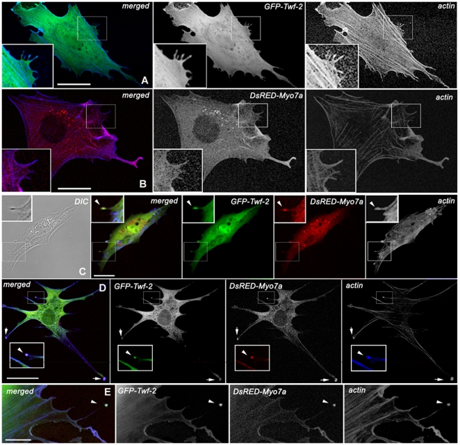
Figure 3. Wild type full-length DsRED-myosinVIIa (red) co-localizes with GFP- twinfilin-2 (green) in filopodia tips.
Confocal images showing distribution of GFP-twinfilin-2, DsRED-myosinVIIa, in BHK-21 cells. Cortical actin was stained with AlexaFluor633/phalloidin (blue). A GFP-twinfilin-2 alone localizes predominantly along the filopodium length. B DsRED-myosinVIIa alone localizes predominantly along the filopodium length. C–E Co-transfection of GFP-Twf-2 and DsRED-Myo7a reveals co-localization of co-expressed proteins at the filopodium tip (arrow heads) and adhesion plaques (arrows) in representative BHK-21 cells. Scale bars: A–H -25 µm.

