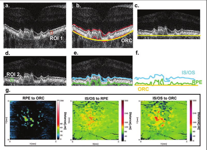
Figure 1.

Retinal layer segmentation procedure. (A) The operator defines the region of interest (ROI 1) inside the required layer. (B) The contour delineating the posterior retina based on ROI 1 is coarsely created and the reference surface, the outer retinal contour (ORC), is calculated. (C) Each cross-sectional image is flattened and cropped with respect to the ORC. (D) The operator defines a new ROI (ROI 2) to accurately segment the posterior retina. (E) Lines representing the basal part of the retinal pigment epithelium (RPE) and the IS/OS junction are automatically identified and three contours corresponding to the RPE, IS/OS, and ORC are calculated. (F) Finally, 200 sets of three contours (the RPE, IS/OS, and ORC) can be analyzed with respect to each other. (G) Contour maps representing the thickness between the RPE and ORC, the RPE and IS/OS, and the ORC and IS/OS.
