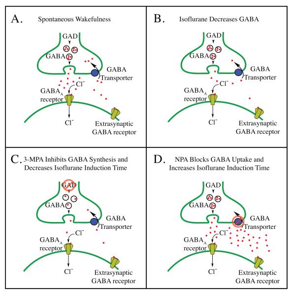
Figure 5.
Schematic illustrating mechanisms of action of isoflurane, 3-mercaptopropionic acid (3-MPA), and nipecotic acid (NPA) within the pontine reticular formation. A. During wakefulness, pontine reticular formation GABA (red dots) is released from vesicles contained in pre-synaptic terminals, increasing chloride ion (Cl-) conductance by activation of post-synaptic and extra-synaptic GABAA receptors. GABA is then actively removed from the synaptic cleft by uptake mechanisms (GABA transporters). B. Isoflurane decreases pontine reticular formation GABA levels, in part, by inhibiting action potential dependent neurotransmitter release,62 by potentiation of GABAA receptor mediated inhibition of GABAergic neurons,15 and/or by pre-synaptic inhibition (disfacilitation) of excitatory inputs acting on GABAergic neurons.35 These three actions of isoflurane on GABAergic transmission likely occur in combinations that vary as a function of brain region. C. Inhibition of glutamic acid decarboxylase (GAD) by 3-MPA decreases GABA levels in the pontine reticular formation, decreases arousal, and facilitates isoflurane-induced loss of wakefulness. D. Inhibition of pontine reticular formation GABA uptake mechanisms by NPA increases GABA levels, increases arousal, and prolongs isoflurane induction time. Parts A — D include extrasynaptic GABA receptors because these receptors are targets for anesthetic drugs,61 and because GABA measured by in vivo microdialysis is thought to act at these extrasynaptic receptors.33

