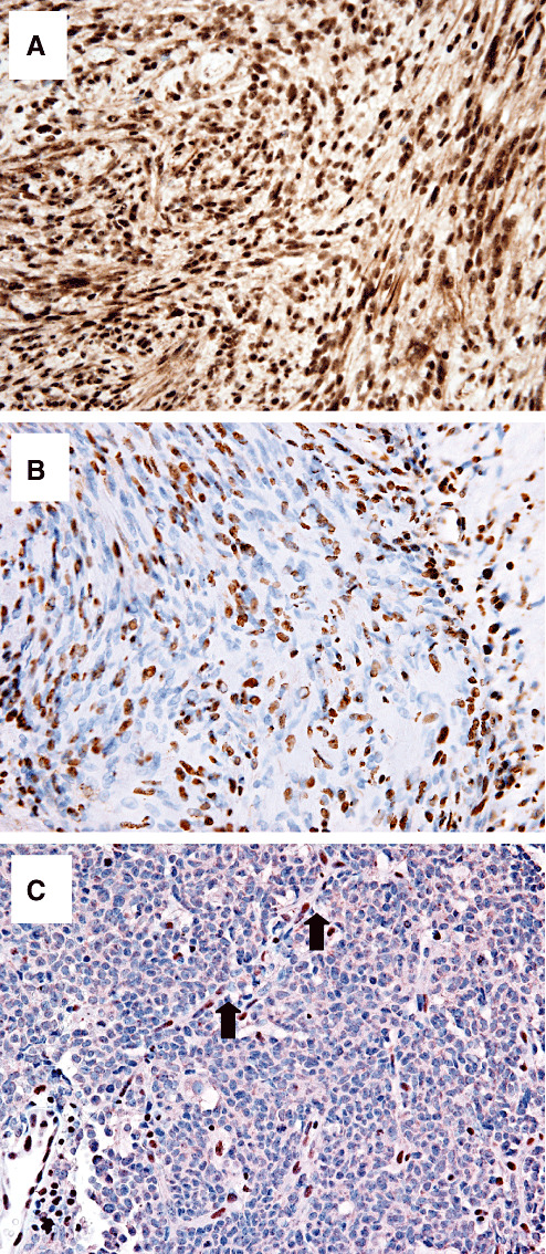
Figure 1.

Immunohistochemical staining of INI1 in schwannomas. A. Solitary, sporadic schwannoma showing diffuse immunopositive staining. B. Schwannoma from a familial schwannomatosis patient showing a mosaic pattern with a subset of immunonegative tumor cells intimately intermixed with immunopositive nuclei. C. Negative control, atypical teratoid/rhabdoid tumor shows diffuse immunonegative tumor; immunopositive capillary endothelial cell nuclei provide an internal positive control (arrows).
