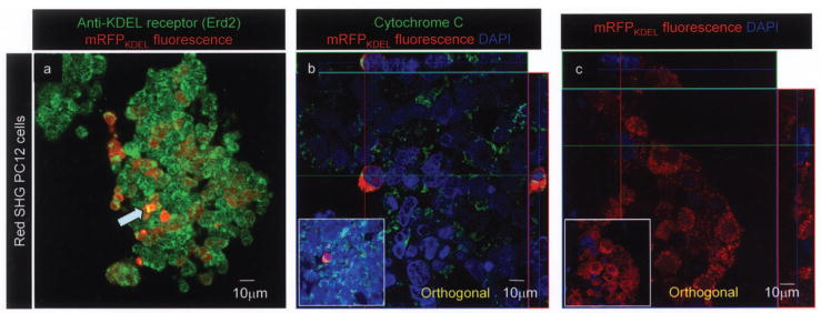
Figure 2.
mRFPKDEL is recognized by Erd2, excluded from the nucleus and mitochondria. Anti-Erd2 antibody staining confirms that the red fluorescence puncta (a) and the punctate green fluorescence from KDEL receptor Erd2 overlap; yellow fluorescence is due to combined fluorescence from Erd2 and mRFPKDEL (a prominent example is indicated by blue arrow). (b) ‘Red SHG mRFPKDEL’ lentivirus infected PC12 cells were stained for cytochrome c; mitochondria appear green, DAPI-stained nuclei are blue. Orthogonal analysis along cytochrome c-stained mitochondria (green on x-axis) does not overlap or include any red puncta (mRFP) that surrounds DAPI-stained blue nuclei. The inset shows normal view of the orthogonal projection. (c) Orthogonal projection shows punctate red fluorescence (red) and the DAPI-stained nucleus (blue) do not overlap. Inset shows the normal view of the field. Original magnification 400×.

