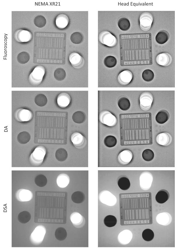
Figure 5.
FP snapshots of the standard 20 cm NEMA XR-21 phantom are shown in the left column, while those for the 15 cm phantom modified by addition of the aluminum filtration are shown in the right column. The top row is acquired using fluoroscopy, middle row Digital Angiography (DA) and the bottom row is acquired using Digital Subtraction Angiography (DSA). (The actual resolution for the bar pattern is not viewable due to reproduction degradation of the images).

