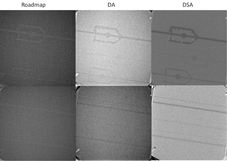
Figure 6.
The results acquired using x-ray imaging of the 15 cm (head-equivalent) phantom containing the 15 mg/cm3 Stenosis/Aneurysm Artery Block 76–705 (top line) and the Low-contrast Artery Insert 76–715 (bottom line) using RoadMap in the first column, Digital Angiograph (DA) middle column, and Digital Subtraction Angiography (DSA) third column

