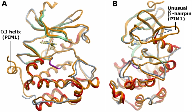
Figure 2. Overall structure of PIM2 and comparison with PIM1.
Overlay of the two proteins (shown in ribbon representation) reveals the strong conservation of the kinase fold. A. PIM1 (2bzh, coloured orange) contains the C-terminal αJ helix that is absent in PIM2 (coloured green for β-strand and red for α-helix). B. The view is rotated by 90° to highlight the unusual β-hairpin in the kinase N-lobe which is partially disordered in the PIM2 structure.

