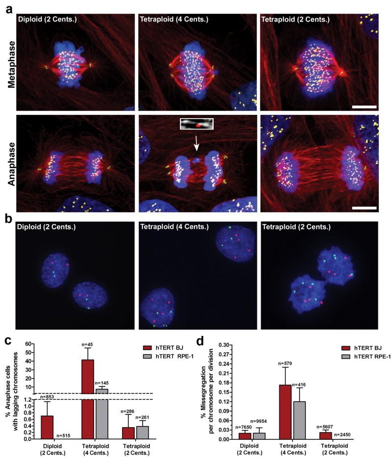
Figure 3. Extra centrosomes promote chromosome missegregation.
a) Human hTERT BJ fibroblasts (Diploid, 2 centrosomes; Tetraploid, 4 centrosomes; Tetraploid, 2 centrosomes) during metaphase and anaphase stained for centrioles (green), microtubules (red), chromosomes (blue), and centromeres (yellow). Arrow indicates a lagging chromosome caused by merotelic attachment (inset; microtubules white, centromere red). b) FISH using centromeric probes specific for chromosomes 6 (green) and 8 (red) in hTERT-BJ fibroblasts. c) Percentage of hTERT BJ (red) and hTERT RPE-1 (grey) cells that exhibit one or more lagging chromosomes during bipolar anaphase (n=number of anaphases counted). d) Missegregation frequency per chromosome per division in hTERT BJ (red) and hTERT RPE-1 (grey) cells (n=number of cell divisions counted). Error bars represent mean ± SE from at least 4 independent experiments. Scale bar, 10 μm.

