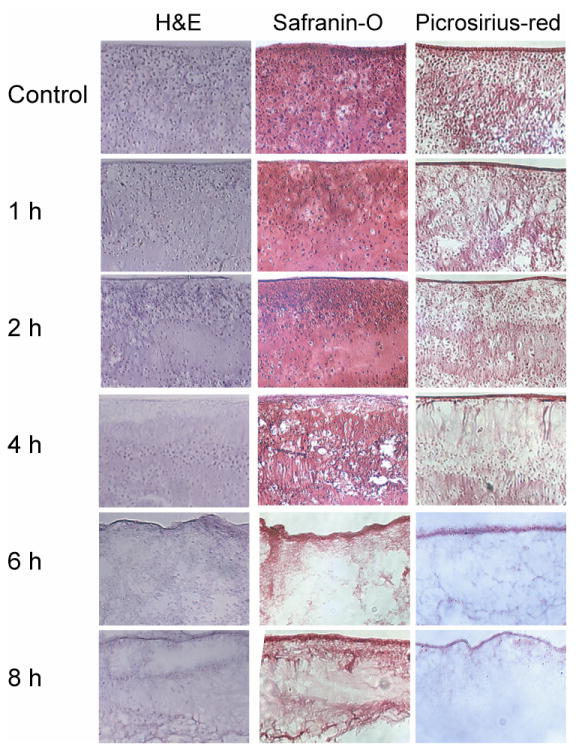
Fig. 2.
Photomicrographs demonstrating construct cellularity, GAG content, and collagen content for treatment groups in phase II. 10× original magnification. Treatment with 2% SDS for 1, 2, and 4 h decreased cellularity while preserving GAG and collagen content, while treatment for 6 and 8 h eliminated all nuclei, but also eliminated GAG and reduced collagen.

