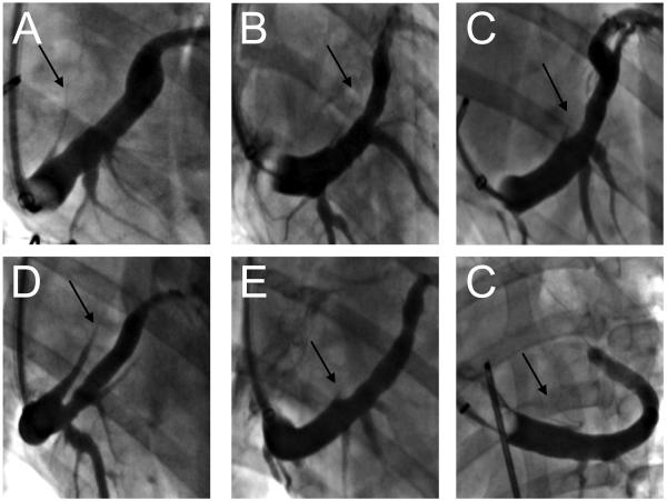
Figure 1.
Angiographic appearance of the VOM in dogs in balloon occlusion venograms. The VOM (black arrows) can be readily identified in the right anterior oblique radiographic projection (A through E) as a posteriorly-directed branch of the coronary sinus. In the left anterior oblique projection (F), it is superiorly directed. Significant variations in caliber and length are obvious.

