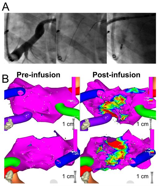
Figure 3.
Posterior left atrial ablation after VOM ethanol infusion. A, Course of the VOM on the coronary sinus venogram (left panel), wire cannulation (mid panel: top wire is in the VOM advanced via a left internal mammary artery angiographic catheter, bottom wire in the main coronary sinus lumen), and selective VOM venogram via an inflated angioplasty balloon. B, Bipolar voltage three-dimensional maps of the left atrial geometry before (left) and after VOM ethanol infusion (right), in the posterior-anterior view (top) and in an inferior view (bottom). The low-voltage area created by VOM ethanol infusion is posteriorly directed, in between left and right pulmonary veins.

