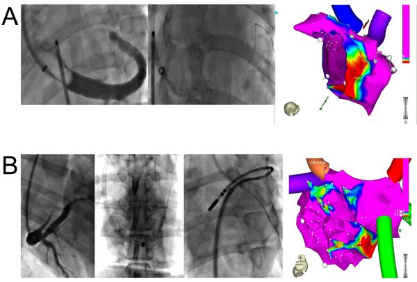
Figure 4.
Anatomical factors leading to reduced ablation area. A, Small VOM. On CS venogram (left panel, left anterior oblique projection), the VOM is a small twig arising from the roof of the coronary sinus. Cannulation was successful, but the angioplasty balloon could only be advanced in the very proximal VOM (mid panel). A small annular area of ablation was created (right panel). B, Communication between VOM and left innominate vein superiorly. Despite a large VOM on the coronary sinus venogram (panel Ba, right anterior oblique projection), and successful VOM cannulation with an angioplasty balloon (panel Bb, left anterior oblique projection), multiple ethanol infusions only created small ablation lesions on voltage maps (panel Bd). A communication between the VOM and the superior vena cava was obvious when the VOM was cannulated from above (panel Bc).

