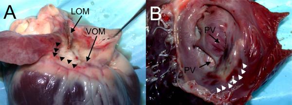
Figure 5.
Ethanol ablation lesions. A, Epicardial aspect, showing the VOM and ligament of Marshall (LOM) areas, with pale discoloration of the ablated areas (arrowheads). B, Endocardial aspect after incision in the left atrial appendage. A pale area of discoloration is shown anterior to the left pulmonary veins (PV), surrounded by small areas of tissue hemorrhage (arrowheads).

