
Figure 1.
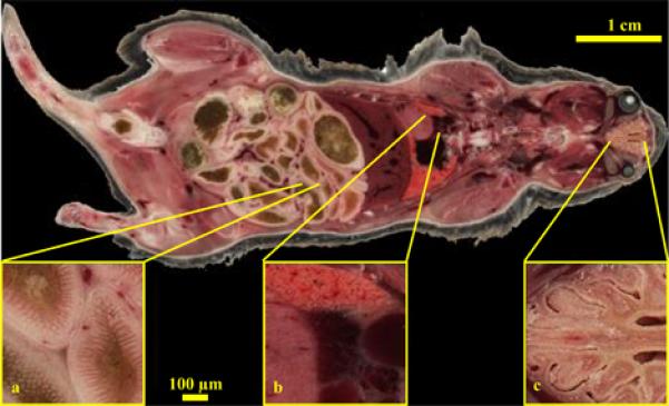
High resolution Cryo-image of a male wild type mouse. A whole mouse was embedded and sectioned and a tiled image acquisition scheme was employed. Each block-face image was composed of 20 tiled images. The low resolution whole image shows all the major organs with remarkable detail like the hepatic vessels of the liver, rectus muscles of the eye, muscle architecture etc. while the high resolution insets show selected areas like the villi of the intestines (a), heart, lung and cardiac vessel architectures (b) and the folds of the nasal septum (c).