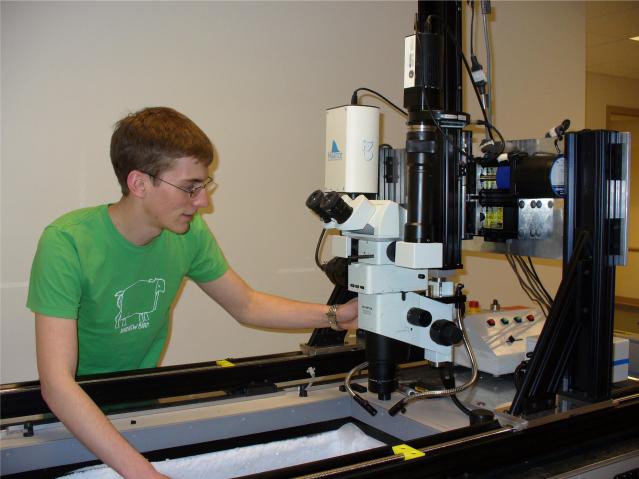
Figure 2.
The present cryo-imaging system. The imaging system comprising of a stereo microscope and digital camera is maneuvered through a xyz robotic positioner. The system is lowered inside the cryo-chamber and positioned above the blockface with the lens parallel to the tissue slicing plane. An imaging workstation computer is interfaced to the cryostat, robotic positioner, illuminators and the camera. Once positioned over the mouse/tissue sample and after imaging protocols are set, the system then automatically goes through a section-and-image sequence without further operator interaction. Both brightfield and fluorescence images in any combination can be captured.

