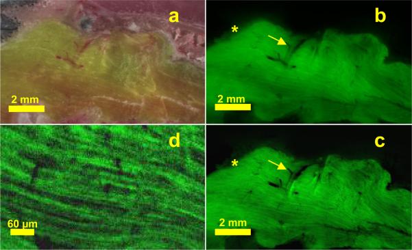
Figure 7.
GFP labeled mouse skeletal muscle, bright field (a), and original (b) and subtraction processed (c) fluorescence cryo-images. In the subtracted image (c), fibers are made apparent due to the removal of subsurface fluorescence. The “halo effect,” has been removed (*), and blood vessels (arrows) have been clarified with removal of subsurface fluorescence. A magnified view of muscle fibers (d) gives single fiber diameter ≈ 10 μm and clearly shows fiber orientation.

