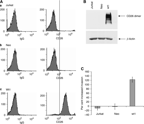
Figure 2.
SDF-1-α-mediated invasion is elevated in Jurkat transfectants expressing CD26. (A) Cell-surface expression of CD26 on Jurkat cell lines. (a) Jurkat-parental, CD26-negative (b) Jurkat-Neo (c) Jurkat-Wt1. Left: cells incubated with PE-labeled isotypic control IgG. Right: cells incubated with PE-labeled anti-CD26 antibody. (B) Expression of CD26 in whole-cell extracts. Equal amounts of protein were run in each lane of a 7.5% gel, transferred to nitrocellulose, and probed with anti-CD26 antibody. Lanes contained Jurkat-parental, Neo cells transfected with empty vector, and wt1 transfectant expressing high levels of CD26, respectively. Note that CD26 runs as a dimer. (C) Invasion assay (as described for Figure 1C). Error bars are shown for the standard error of the mean.

