Figure 1.

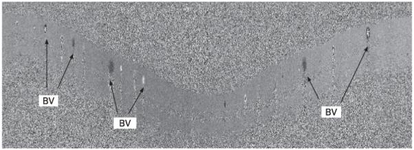
Doppler optical coherence tomography image. The 2.0 mm radius cylindrical section of the retinal is unwrapped, and the Doppler shift is shown in grey scale. BV, branch vein.

Doppler optical coherence tomography image. The 2.0 mm radius cylindrical section of the retinal is unwrapped, and the Doppler shift is shown in grey scale. BV, branch vein.