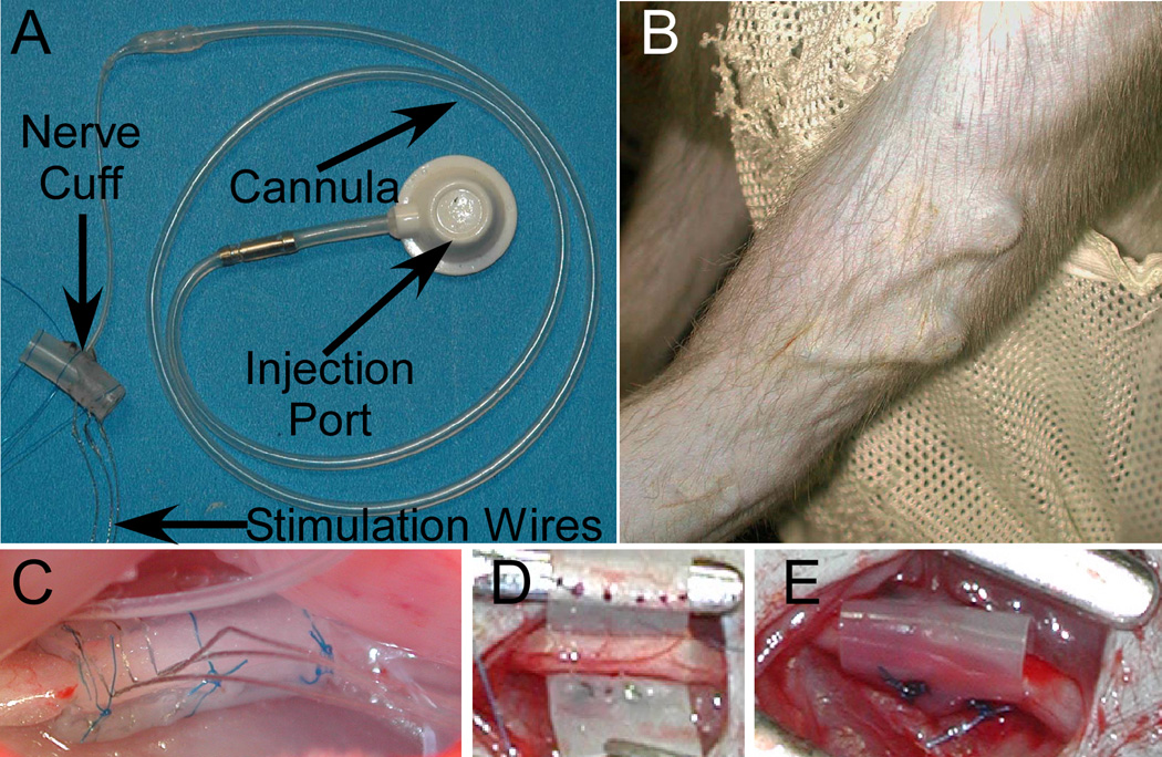
Figure 1.
A: Nerve cuff, cannula, and injection port used in rabbit experiments. B: The appearance of the injection dome beneath the skin in the upper arm of monkey M2. C: A nerve cuff around the rabbit sciatic nerve. The 3 stimulating wires are visible in the foreground. D-E show the implantation of a cuff around the ulnar nerve in M2 before (D) and after (E) the silastic has been shaped into a tube around the nerve.

