
Fig. 2.
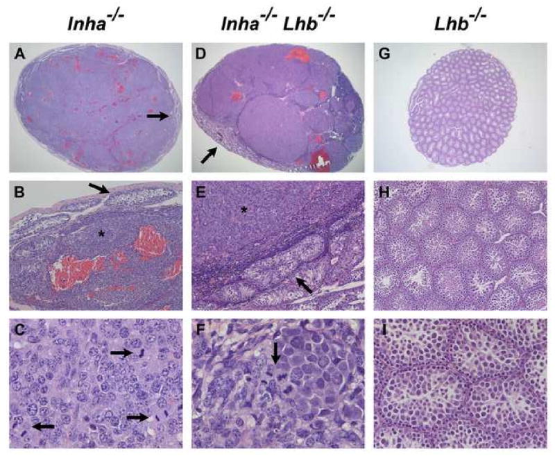
Histological analysis of testicular tumors from end-stage Inha-/- and Inha-/-Lhb-/- males as compared to testes from 12-week-old Lhb-/- males (n ≥ 5 mice of each sex and genotype). Multiple areas of hemorrhage are present in testicular tumors from Inha-/- (A) and Inha-/-Lhb-/- (D) mutants. The tumors display poorly differentiated neoplastic cells (asterisks in B, E) that are compressing the few remaining seminiferous tubules (arrows in A, B, D, E). Numerous mitotic figures are also present (arrows in C, F). In contrast, Lhb-/- testes are non-hemorrhagic and do not contain tumorigenic foci (G-I). The major histologic abnormalities in Lhb-/- testes are their small size (G), sparse interstitium with few Leydig cells (H), and spermatogenic arrest at the round spermatid stage (I), consistent with prior observations (Ma et al., 2004). Ages of mice: A-C: 9 wks; D-F: 18 wks; G-I: 12 wks. Magnification: A: ×15.6; B, E, H: ×100; C, F: ×640; D: ×12.5; G: ×20; I: ×250.