
Fig. 3.
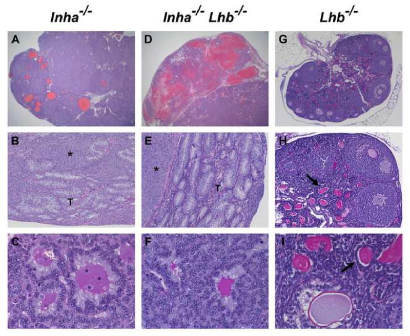
Histological analysis of ovarian tumors from end-stage Inha-/- and Inha-/-Lhb-/- females as compared to ovaries from 12-week-old Lhb-/- females (n ≥ 5 mice of each sex and genotype). The ovarian tumors from both Inha-/- and Inha-/-Lhb-/- mutants are hemorrhagic (A, D) and composed of poorly differentiated neoplastic cells (asterisks in B, E) along with tubular structures (T) lined with Sertoli-like cells and Call-Exner bodies (C, F) that are characteristic of granulosa cell tumors. In contrast, Lhb-/- ovaries are non-hemorrhagic and do not contain tumorigenic foci (G-I). These ovaries are small and contain numerous degenerating oocytes (arrows in H, I). Preovulatory follicles and corpora lutea are notably absent from Lhb-/- ovaries, indicating a block in folliculogenesis at the antral follicle stage, as previously reported (Ma et al., 2004). Ages of mice: A: 12 wks; B: 25 wks; C: 15 wks; D: 14 wks; E: 23 wks; F: 13 wks; G-I: 12 wks. Magnification: A, D: ×12.5; B, E, H: ×100; C, F, I: ×400; G: ×40.