
Fig. 5.
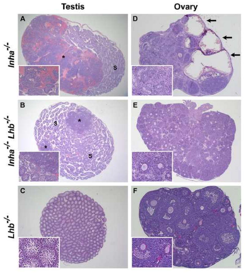
Histological analysis of testes (A-C) and ovaries (D-F) from 6-week-old Inha-/-, Inha-/-Lhb-/-, and Lhb-/- mice (n ≥ 5 gonads of each sex and genotype). (A) Inha-/- testis predominantly containing tumor cells and hemorrhage (asterisk), with a smaller region of normal seminiferous tubules (S). In contrast, the testis of an age-matched Inha-/-Lhb-/- male (B) is filled with seminiferous tubules, and only two small tumor foci (asterisks) are readily apparent. (C) No tumor foci are present in Lhb-/- testes. (D) Inha-/- ovary with architecture almost entirely disrupted by numerous hemorrhagic cysts (arrows) and nodules of poorly differentiated neoplastic cells. In contrast, the ovaries from age-matched Inha-/-Lhb-/- (E) and Lhb-/- (F) females are non-hemorrhagic, intact, and contain multiple oocytes. Magnification: A: ×12.5; B: ×15.6; C: ×20; D: ×31.3; E: ×25; F: ×62.5.