
Fig. 6.
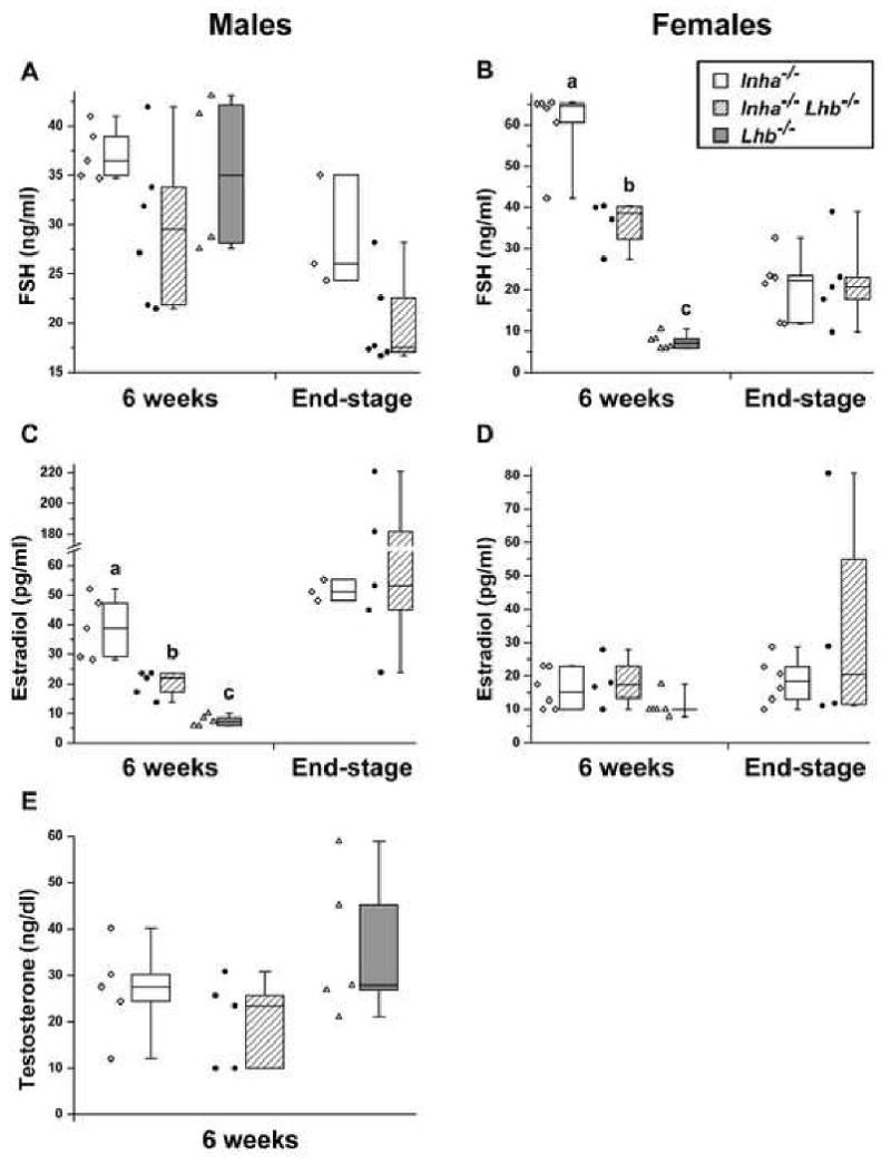
Serum FSH (A, B), estradiol (C, D), and testosterone (E) measurements for Inha-/-, Inha-/-Lhb-/-, and Lhb-/- males and females. The individual data points are shown and are also represented as standard box plots with whiskers extending to the minimum and maximum values. In 6-week-old females, FSH is significantly different between all three mutants, while in 6-week-old males, estradiol is significantly different between all three mutants (p<0.0001, oneway ANOVA followed by Tukey's HSD test). In addition, both 6-week-old and end-stage Inha-/-Lhb-/- males demonstrate a trend toward lower FSH compared to Inha-/- controls. Ages of end-stage Inha-/-mice: A, C: 9-11 wks; B, D: 10-25 wks. Ages of end-stage Inha-/- Lhb-/- mice: A, C: 7-60 wks; B, D: 13-33 wks.