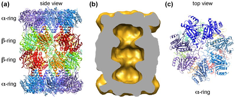
Figure 1.
Architecture of the 20S CP. (a): A ribbon diagram of the yeast 20S proteasome (pdb accession code: 1RYP) [4–6]. Two inner β-rings and two outer α-rings are marked. (b) Volume of the yeast 20S CP, calculated from atomic coordinates and filtered to 20 Å. The volume is cut in half, showing three chambers within the 20S CP. (c) The top surface of the α-ring. The N-termini of the α-subunits form a closed gate block the entrance to the inner chamber of the 20S CP.

