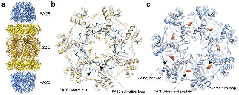
Figure 2.
Gate opening of the 20S CP by proteasomal activators. (a) Ribbon diagram of 20S-PA26 complex (PDB accession code: 1YA7) [17]. (b) Top view of the 20S CP’s α-ring, showing both a closed gate (light gray, PDB accession code: 3C92) and an open gate (yellow) in the presence of the PA26 (blue). The locations of the PA26 C-terminus and activation loop are marked. Notice that the 20S reverse turn loop (pointed in (c)) is moved by the PA26 activation loop. (c) CryoEM structure of 20S-PAN peptides, with an open-gate conformation (light blue, PDB accession code: 3C91). The structure of a closed gate is shown in light gray (PDB access code: 3C92) [33]. The densities correspond to the PAN C-terminal peptide, indicates the binding site of PAN’s C-termini in the 20S CP.

