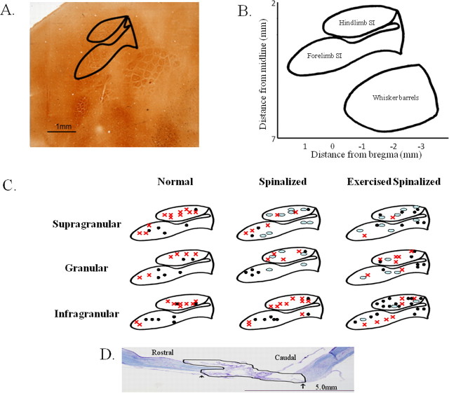
Figure 1.
Penetration sites for the acute single-neuron mapping of forelimb and hindlimb somatosensory cortex in normal, spinalized, and exercised spinalized rats. A, The flattened somatosensory cortex, when stained with cytochrome oxidase in a normal adult rat, reveals the locations that we targeted in this study. Solid lines outline the hindlimb and forelimb somatosensory cortices. B, Schematic of the flattened somatosensory cortex based on 1 mm grids showing the stereotaxic coordinates of the forelimb and hindlimb somatosensory cortex. C, Penetration sites into the forelimb and hindlimb somatosensory cortex for each group of rats. Responses were recorded from electrodes placed in the supragranular, granular, and infragranular layers of the hindlimb and forelimb somatosensory cortices. Black circles, Electrode penetration sites that elicited cells which responded to sensory stimulation. Red x, Electrode penetration sites that elicited cells which did not respond to sensory stimulation. Ovals, Electrode penetration sites that did not elicit any cells. D, Nissl/myelin staining of the spinal cord of a representative adult rat transected as a neonate at the T8/T9 level. No cell bodies or axons were observed in the transection site (outlined in black). The extent of this representative injury was comparable with the other injuries. The average rostral to caudal extent of the injury was 6.4 ± 2.2 mm, and no differences were observed in the extent of the injury between the spinalized and exercised spinalized groups. Scale bar, 5.0 mm.

