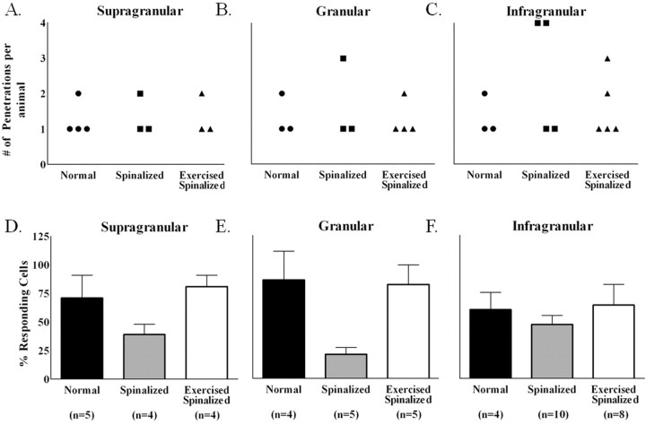
Figure 5.
Exercise restored the percentage of cells that responded to sensory stimulations in the forelimb somatosensory cortex of spinalized rats. Data were collected from four normal, four spinalized, and five exercised spinalized rats. A–C, The numbers of electrode penetrations within the forelimb somatosensory cortex from which cells were detected are shown for each animal (circles represent normal animals, squares represent spinalized animals, and triangles represent exercised spinalized animals). D–F, The averages of the percentages of cells that responded to sensory stimuli for each penetration (n = total number of penetrations) were plotted as a per layer basis for each animal group. As expected, a high percentage of cells in all cortical layers of the forelimb cortex responded to sensory stimulation of the forelimb [supragranular (71% of 42 cells), granular (86% of 21 cells), and infragranular (60% of 77 cells)]. Neonatal spinalization decreased the percentages of sensory responsive cells in all layers of the forelimb cortex [supragranular (39% of 31 cells), granular (21% of 28 cells), and infragranular (47% of 181 cells)] when compared with those of the normal rats. Exercise restored the percentages of sensory responsive cells in all cortical layers of spinalized rats, comparable with those of normals [supragranular (81% of 21 cells), granular (82% of 27 cells), and infragranular (64% of 90 cells)]. Error bars indicate SEM.

