
Figure 3.
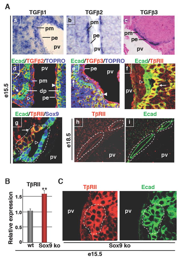
TGFβ signaling and development of bile ducts in wild-type and SOX9-deficient livers. (A) In situ hybridization showing wide expression of TGFβ1 in the liver, while TGFβ2 and TGFβ3 mRNAs were predominantly found in the portal mesenchyme (a–c). Immunostaining showed binding of TGFβ2 and TGFβ3 to the parenchymal side of PDS (d, e). Immunostaining showing expression of TβRII on the parenchymal side of PDS and lack of expression in mature ducts (f–i). Arrows: TGFβ2, TGFβ3 or TβRII staining on parenchymal side of PDS; Arrowheads: TGFβ2 or TGFβ3 staining on single-layered ductal plate; open arrowheads: lack of TGFβ2, TGFβ3 or TβrII staining on portal side of PDS. (B) Q-PCR showing overexpression of TβRII in e15.5 Alfp-Cre-Sox9loxP/loxP (Sox9 ko) livers. (C) Immunostainings showed that primitive ductal structures detected by E-cadherin (E-cad) labeling expressed TβRII on both the portal side and parenchymal side in Alfp-Cre-Sox9loxP/loxP livers, in contrast to wild-type primitive ductal structures which express TβRII only on the parenchymal side (see panels Af, g). dp, single-layered ductal plate; pe, portal endothelium; pm, portal mesenchyme; pv, portal vein; *, lumens of developing bile ducts.