
Figure 1.
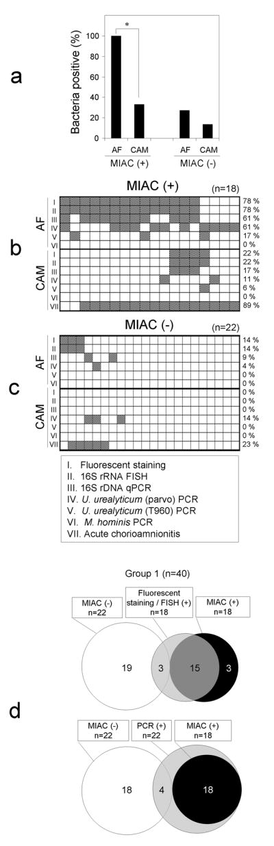
Comparison of Group 1 cases without (n=22) and with (n=18) MIAC defined by positive amniotic fluid culture. (a) Proportion of the cases in which bacterial traits were demonstrated by any fluorescent staining, FISH, 16S rDNA qPCR, or real-time PCR for U. urealyticum or M. hominis in the amniotic fluid and in the chorioamniotic membranes. Bacteria were more frequently detected in the amniotic fluid than in the chorioamniotic membranes for cases with MIAC (*P<0.0001). (b, c) Maps showing proportion of the cases with histologic chorioamnionitis or the cases in which bacteria were detected in amniotic fluid and chorioamniotic membranes according to assay methods employed. (d) Venn diagrams showing proportion of cases in which bacteria were detected by different techniques. White and black circles represent cases without and with MIAC. Microscopic examinations (fluorescent staining/FISH) or PCR (16S rDNA qPCR, or real-time PCR for U. urealyticum or M. hominis) are in gray circles, for which the number represents the cases positive for bacteria in the amniotic fluid or in the chorioamniotic membranes. MIAC: microbial invasion of the amniotic cavity, AF: amniotic fluid, CAM: chorioamniotic membranes.