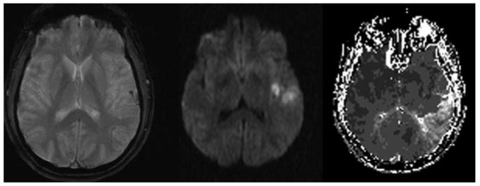
Figure.
Distal left MCAO in the setting of acute stroke. GRE sequence (left) reveals blooming artifact associated with thrombus in a cortical arterial branch. DWI (middle) shows ischemic changes approximating the ischemic core in a limited region of tissue immediately beyond the MCAO. Time-to-peak perfusion MRI sequence (right) illustrates the delayed flow of collateral perfusion to a far more extensive region of penumbra.

