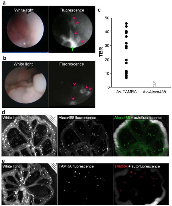
Figure 9.
In vivo fluorescence endoscopic images in tumor bearing mice enhanced by Av-Alexa488 (a) and Av-TAMRA (b). The pink arrow heads show the tumor nodules. The tumors were clearly visualized with the activatable probe, Av-TAMRA. In contrast, Av-Alexa488, an always-on probe, showed high background signal and high fluorescence from excess injectate in the peritoneal cavity (green arrow). The measured tumor-to-background ratio (TBR) was higher for Av-TAMRA than Av-Alexa488 (c). Fluorescence spectral images of the peritoneal membranes for Av-Alexa488 (d) and Av-TAMRA (e). The results were consistent with endoscopic images. Tumors were detected with low background signal for Av-TAMRA, but the background was high for Av-Alexa488.

