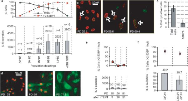
Figure 2. Telomeric PDDF promotes increased IL-6 secretion.

(a) HCA2 populations were cultured until replicative senescence (PD>71). Top panel: Populations were sampled at the indicated PDs (lower panel X axis). Single cells were analyzed for DNA synthesis (BrdU incorporation over 24 h) and 53BP1 foci (cells with 3 or more foci). Lower panel: Conditioned media were collected over a 24 h interval from the populations analyzed in the top panel. IL-6 was measured by ELISA, and is reported as 10−6 pg/cell/day (n= number of cell populations analyzed).
(b) HCA2 cells at the indicated PDs were fixed and single cells analyzed simultaneously for 53BP1 foci (green) and BrdU incorporation over a 24 h interval (red). Arrows mark nuclei that simultaneously harbor 53BP1 foci and BrdU staining.
(c) Cells at PD35 were pulsed with BrdU for 1 h and fixed. Single cells were analyzed simultaneously for 53BP1 foci and BrdU incorporation. BrdU incorporation was scored independent of 53BP1 foci (Total cells) or only in cells with 53BP1 foci (53BP1+).
(d) Cells at the indicated PD levels were pulsed with BrdU for 24 h and fixed. Single cells were analyzed by immunofluorescence for intracellular IL-6 (green) and BrdU incorporation (red).
(e) Cells were infected with a retrovirus expressing hTERT, selected and cultured for the indicated doublings (PD after hTERT). At the indicated PDs, cells were analyzed for 3 or more 53BP1 foci (top panels) or IL-6 secretion (lower panels), as described for Fig. 2a.
(f) Early passage HCA2 or HCA2 cells expressing hTERT were irradiated with 10 Gy, allowed to recover for 8 d, and analyzed for 53BP1 foci (top panel) and IL-6 secretion as described for Fig. 2a. IL-6 secretion is reported as the fold increase compared to unirradiated control cells. There was a non-significant reduction in 53BP1 foci and slight reduction in IL-6 levels (p=0.036, two-tailed student T-test for unpaired samples) in hTERT cells.
