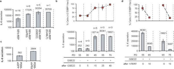
Figure 3. Loss of p53 accelerates PPDF formation and IL-6 secretion.

(a) Replicatively senescent HCA2 cells were infected with the indicated lentiviruses and IL-6 secretion was analyzed 3−4 PDs following reversal of the senescence arrest. IL-6 secretion is reported as 10−6 pg/cell/day on a log scale (n= number of cell populations analyzed).
(b) Early passage HCA2 cells were infected with a GSE22-expressing retrovirus, selected for 4 d (∼3 PDs) and IL-6 secretion was measured ∼2 PDs later. Cells were cultured for the indicated PDs and assessed for 53BP1 foci (top panel) and IL-6 secretion (lower panel) (10−6 pg/cell/day on a log scale) (n= number of cell populations analyzed). Because GSE22 prevents p53 tetramerization, which is required for transactivation and rapid degradation, GSE22-expressing cells contained abundant inactive p53 protein (see Supplementary Information, Fig. S3a).
(c) Early passage HCA2 cells (PD35) were infected with lentiviruses expressing either shp53 or eGFP, selected and cultured until PD55, and analyzed for IL-6 secretion (10−6 pg/cell/day on a log scale).
(d) Two HCA2-GSE22 populations were infected with a retrovirus expressing hTERT and analyzed for 53BP1 foci and IL-6 secretion as described above.
