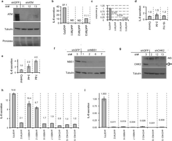
Figure 4. DDR signaling is required for the cytokine response to PDDF.

(a) HCA2 cells were infected with lentiviruses expressing shRNAs against GFP (control, shGFP3) or ATM (shATM-11, −12, or −13) and allowed to recover for 5 d. Whole cell lysates were analyzed by western blotting for the indicated proteins. Ponceau staining shows the total proteins.
(b) HCA2 cells were infected as above and irradiated with 10 Gy X-Ray. After 9 d, CM were collected over 24 h and assessed for IL-6 by ELISA. IL-6 secretion is reported as fold change over unirradiated shGFP control. ND=not detected.
(c) Replicatively senescent HCA2 were infected as above and allowed to recover for 6 d. Conditioned media CM were collected over 24 h and assessed for IL-6 secretion (reported as fold change over shGFP control).
(d) A-T cells were irradiated with 10 Gy and allowed to recover for 2 or 10 d. CM were collected over 24 h, and analyzed for IL-6 by ELISA (reported as fold increase compared to unirradiated A-T cells; numerical values are given above the bars).
(e) Seckel syndrome cells were irradiated and analyzed for IL-6 as described above for A-T cells. (f-g) HCA2 cells were infected with lentiviruses expressing shRNAs against GFP (control, shGFP3), NBS1 (shNBS1−1, −2, −6, or −7)) or CHK2 (shCHK2−2, −12, or −13), selected and allowed to recover for 7 d. Whole cell lysates were analyzed by western blotting for the indicated proteins. The arrow indicates CHK2; NS indicates a non-specific band detected by the antibody.
(h) HCA2 cells from (Fig4f-g) with the indicated shRNA-expressing lentiviruses were irradiated with 10 Gy X-Ray. After 2 d, CM were collected over 24 h and assessed for IL-6 using ELISA (reported as fold change over unirradiated shGFP control; numerical values are given above the bars).
(i) Replicatively senescent HCA2 cells were infected with indicated shRNA-expressing lentiviruses and allowed to recover for 6 d. CM were collected over 24 h and assessed for IL-6(reported as fold change over shGFP control; numerical values are given above the bars).
